Changing Lives One Smile At A Time!

Radiology

Radiology

In Dental Studio, we have established a spacious environment and all other conditions for fellow dentists and their patients to perform X-rays using the most sophisticated and safest X-ray machine of the leading global brand, KAVO.

Senior radiology technicians, with continuous education and vast experience in dental radiology, are in charge of performing X-rays.

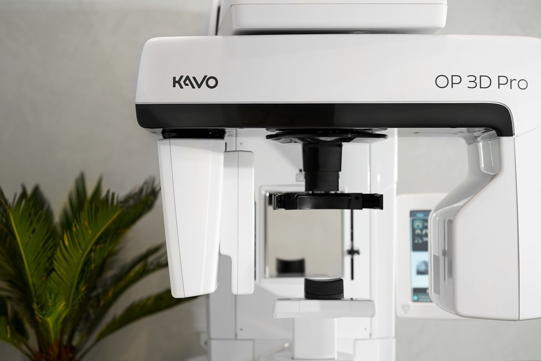
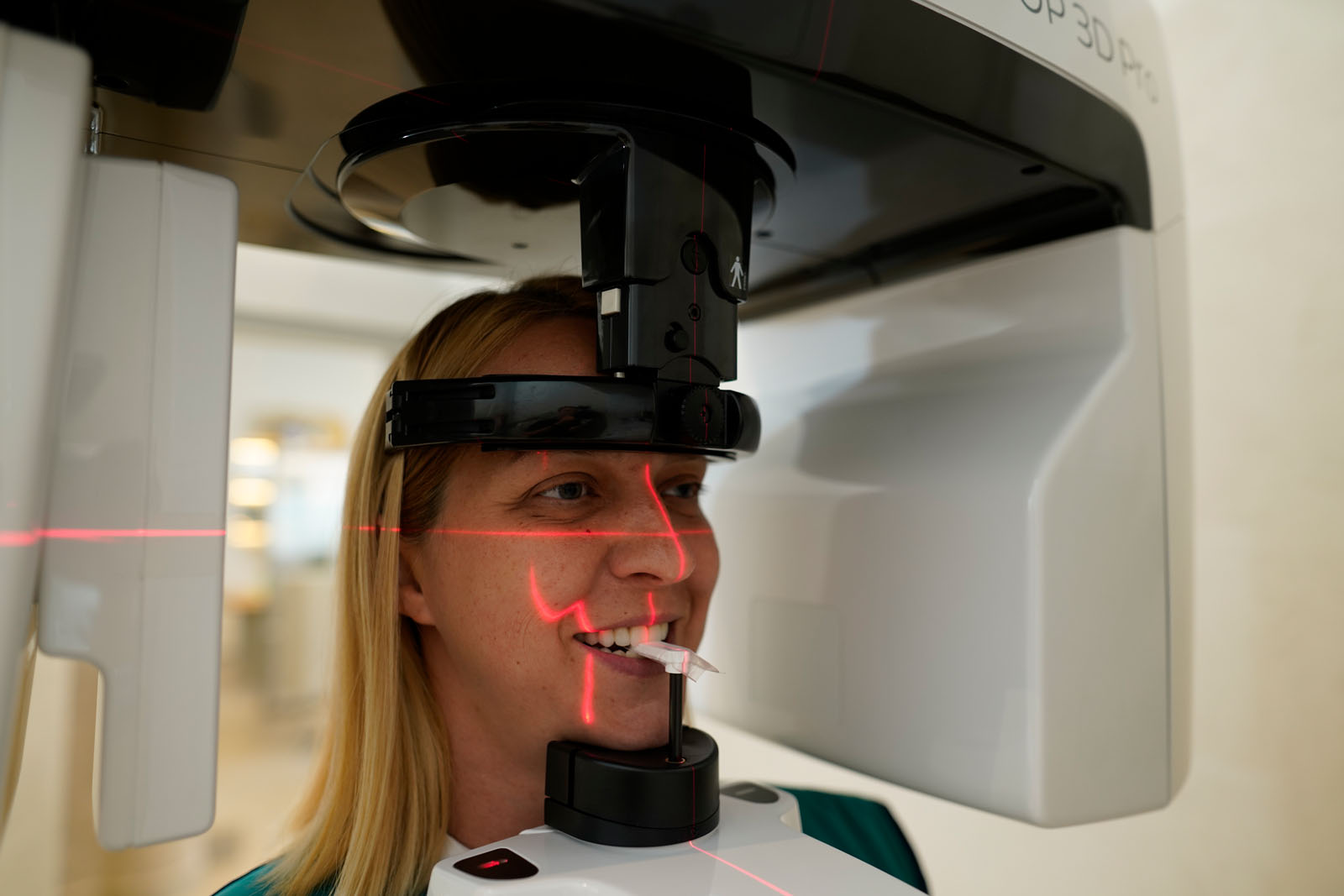
3D radiogram - CBCT (cone beam computered tomography)

3D radiogram - CBCT (cone beam computed tomography) represents dental cone beam computed tomography which, in addition to producing X-rays, also applies a math-based image processing method. This digital geometrical processing is used for generating three-dimensional images of the inside of an object, consisting of a large series of two-dimensional images produced during one rotation of the gantry around its axis. 
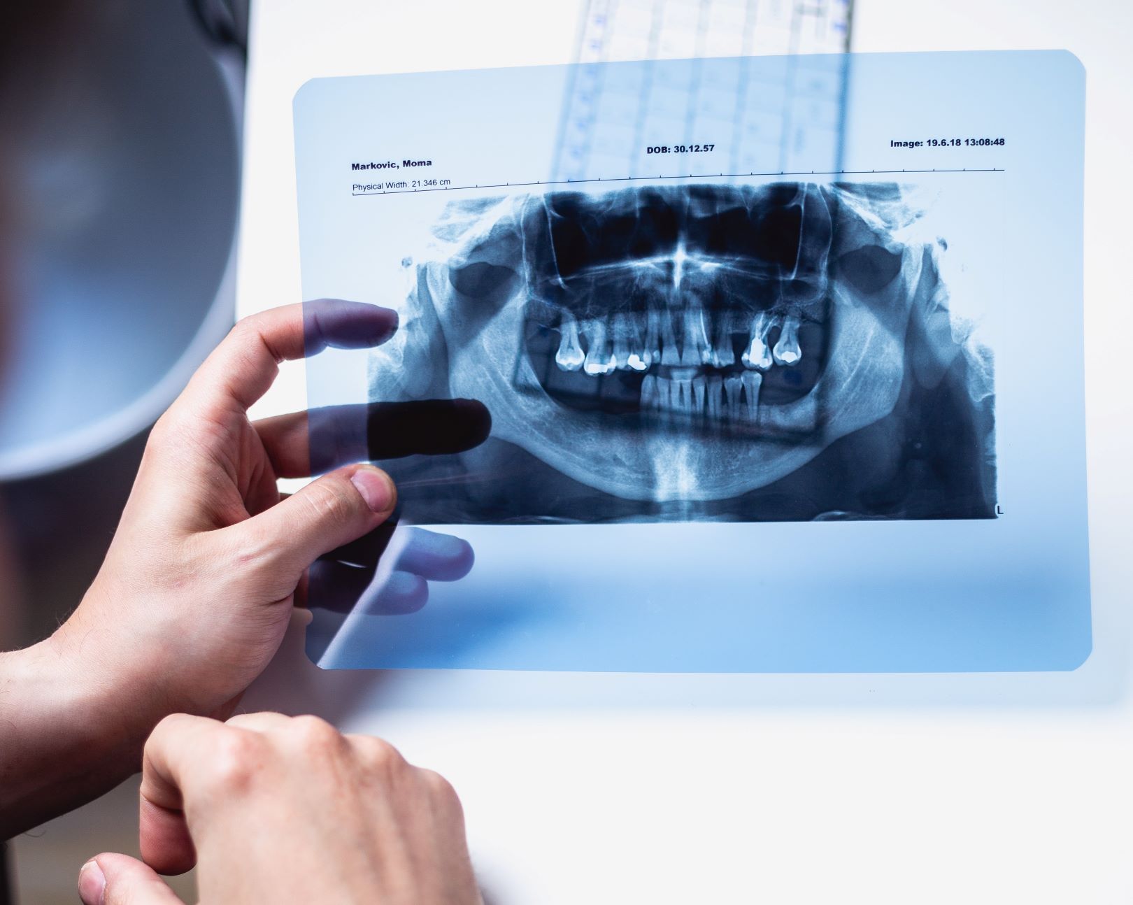
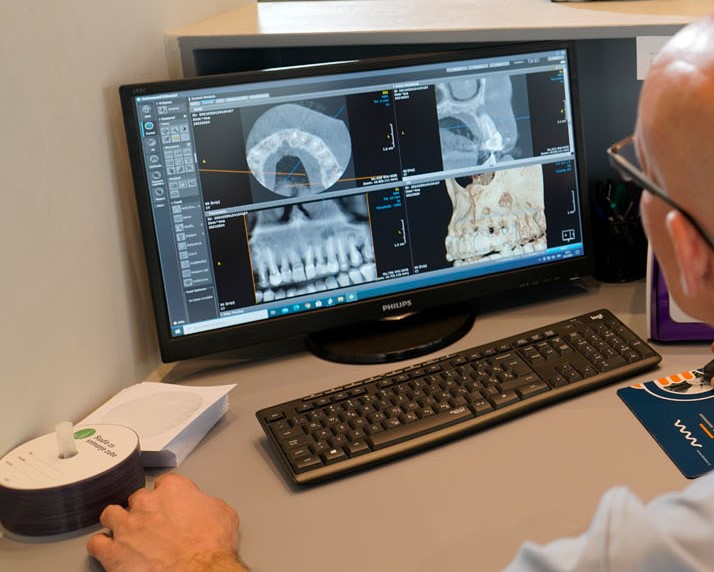
Such X-ray view enables dentists to define a diagnosis and therapy plan more easily and precisely, with an ultimate goal of successful treatment. It provides more planes (axial, panoramic, sagittal), as well as an insight into details which are not visible in standard two-dimensional images. Four different field sizes (5 x 5, 6 x 8, 8 x 8, 8 x 15) enable imaging of regions of interest with minimal radiation dosages.

Digital orthopan

Digital orthopan is considered the basic radiological image, which also represents the main prerequisite for all complex dental therapy procedures. Orthopan is a panoramic image of all teeth, maxillary sinuses, temporomandibular joints and bone tissue of the maxilla and mandible. In our diagnostics centre, digital orthopan is issued on high-resolution foil or sent in digital form, via email or on a CD.

Digital orthopan for children

Digital orthopan for children – Digital orthopan with a reduced radiation dosage is produced for children especially and it represents maximum safety. It is an image with a shorter exposition time and lower radiation dosages (50% less radiation).

Retro-alveolar X-ray

Retro-alveolar X-ray – Intraoral X-ray is a precise image indicating specific details and it has been used vastly. It is used for a very small area, most often for disorder or disease of an individual tooth. In our diagnostics centre, there is an option of obtaining an enlarged retro-alveolar X-ray processed from several angles, on a large high-resolution foil, for an easier diagnostic procedure. As with other types of X-rays, a retro-alveolar one can be obtained in electronic form.

Digital X-rays of temporomandibular joints

Digital X-rays of temporomandibular joints are images that are used to easily diagnose parafunctions (such as teeth grinding and jaw clenching, for instance) or structural changes on a temporomandibular joint, which attaches the mandible to the skull bones. A temporomandibular joint X-ray is produced in two projections, with open and closed mouth, namely in an antero-posterior projection.

Scanogram

Scanogram – Segmental orthopantomographic X-ray is an image of an individual area of interest for a dentist. In producing this type of image, only an area of interest is exposed to X-ray, rather than both jaws. Digital scanograms can include each quadrant individually, with a lower radiation dosage, and the information obtained from such images is comprehensive and precise.

Bitewing technique of dental X-raying

Bitewing technique of dental X-raying is a special technique of extraoral imaging where teeth crowns of the maxilla and mandible are visible. This imaging technique is used to precisely diagnose cavities on lateral-approximal teeth surfaces, which, generally, represent one of the most common diagnostics procedures for a dentist, as well as one of the most frequent reasons why young patients visit a dentist.

Make your dream smile a reality!

Call us or book your appointment today